
Figure 1.
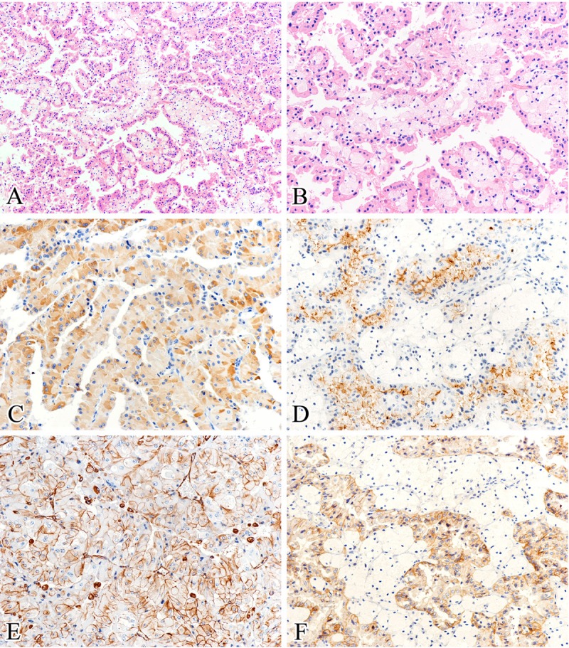
Histological and immunohistochemical features. A: All tumors showed a predominant papillary structure and delicate fibrovascular cores, with foamy macrophages usually seen in the papilla cores (hematoxylin and eosin). B: Papillae were lined by single layers of tumor cells with large, deeply eosinophilic and finely granular cytoplasm, and low grade nuclei (hematoxylin and eosin). C-F: All cases exhibited strong, diffuse positive for racemase (C), CD10 (D), vimentin (E), and MET (F) staining (immunohistochemical staining). (Original magnifications: A × 100; B-F × 200).