
Figure 3.
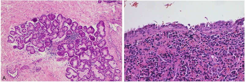
Microscopic features. A: Salivary tissue contained in the cyst wall (HE, x100). B: Ciliated epithelial lining of the Rathke cleft cyst abutting on adenohypophyseal tissue (HE, x200).
Official websites use .gov
A
.gov website belongs to an official
government organization in the United States.
Secure .gov websites use HTTPS
A lock (
) or https:// means you've safely
connected to the .gov website. Share sensitive
information only on official, secure websites.
Microscopic features. A: Salivary tissue contained in the cyst wall (HE, x100). B: Ciliated epithelial lining of the Rathke cleft cyst abutting on adenohypophyseal tissue (HE, x200).