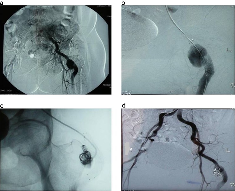
Fig. 3.
Digital subtraction angiography. A broad-based saccular pseudoaneurysm of persistent sciatic artery (a), placement of a metallic stent in the artery (b), obstruction of pseudoaneurysm with spiral coils (c), control angiography that shows the aneurysm is out of vascular tract and perfusion of the lower extremity is completely normal (d)

