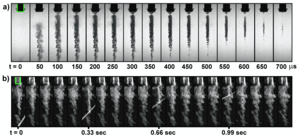
Figure 2.

Representative image sequences of cavitation during stone comminution experiments in free field (PRF = 3 Hz) without the jet. The shadowgraph high-speed image series (a) were captured after the 20th shocks using a 10 μs exposure time. The B-mode ultrasound images (b) were recorded during the experiment (around the 200th shock) to observe nuclei persistence during subsequent shockwave exposures. Bright rays seen on several B-mode image frames correspond to the interference produced by the release of lithotripter shock wave. The lifetime of detectable bubble nuclei is ~7 seconds. The stone holder ∅out = 17 mm) is highlighted in the first frame.
