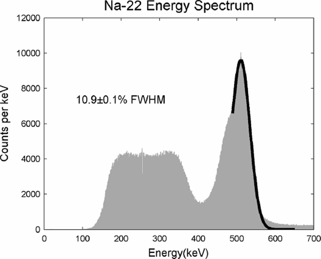
Fig. 13.
An energy spectrum from an 8 × 8 array of l×l×l mm LSO-PSAPD detector. The gain is calibrated per crystal and the resulting energies combined to form the spectrum of the whole detector. The energy resolution is l0.9±0.1% FWHM and the lutetium X-ray escape energies are visible in the lower energy side of the photopeak.

