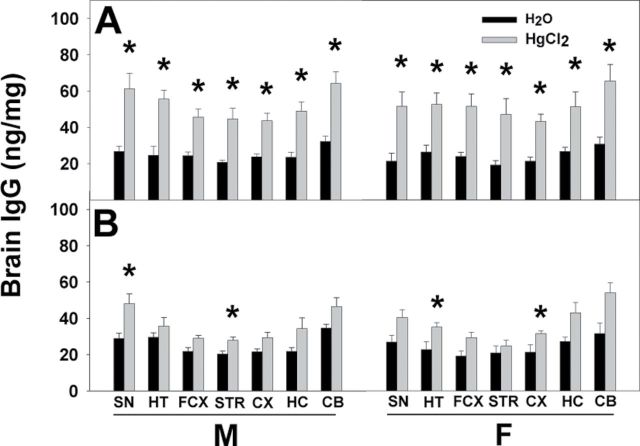
Fig. 3.
IgG levels in brain regions of pnd21 offspring. Whole brains of pnd21 SFvF1 (A) and FvSF1 (B) offspring were dissected into SN, HT, FCX, STR, CX, HC, and CB, and homogenates of each region were used to assess presence of IgG by ELISA. GAM IgG γ-chain Abs were used as capture Abs, and peroxidase-conjugated GAM IgG whole molecule Abs were used as detection Abs for quantification of brain IgG (ng/mg protein). M, male; F, female; * indicates a significant difference of the Hg group compared with the counterpart water group (p < 0.05). Number of mice used (H2O; Hg): M (5; 7) and F (5; 7) SFvF1, M (7; 6) and F (4; 6) FvSF1.

