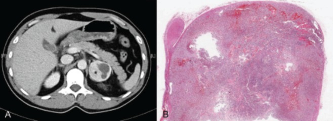
Figure 1. Computed Tomography and Histological Images of the Pheochromocytoma.

A: Abdominal computed tomography showing a left adrenal mass of 50 mm in diameter with rounded, well-defined edges, and hyperdense areas of cystic necrosis inside (asterisk). B: Histological panoramic view of the pheochromocitoma. On the left side of the picture there is a normal adrenal gland on which sits the tumor with a large nodule with areas of hemorrhagic aspect, especially in the tumor periphery.
