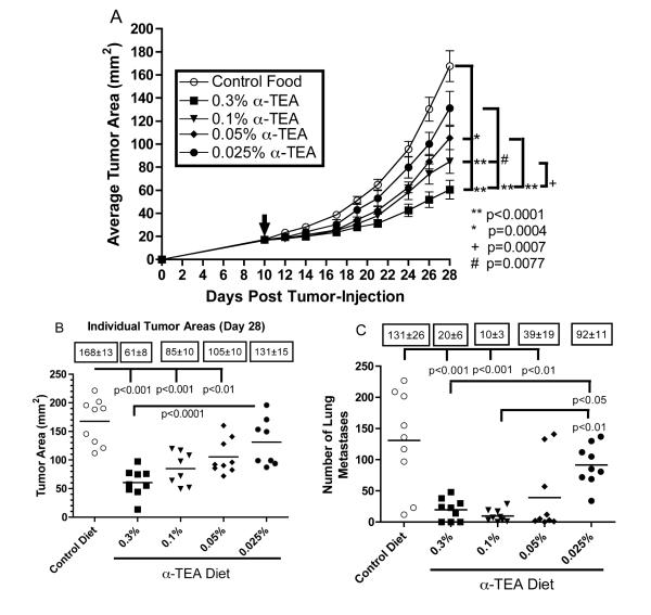
Figure 1. Effect of dietary delivery of α-TEA on primary tumor growth.
BALB/c mice were injected with 4T1 mammary tumor cells in the right mammary fat pad (day 0). When tumors became palpable ( ,~17 mm2, day 10 post-tumor cell injection), mice received α-TEA in the diet for 18 days. Untreated mice received control diet throughout the study. (A) The values represent the mean tumor areas ± SEM of 9 mice per group. To compare tumor growth rates, growth curves were transformed to linearity and linear regression analysis was used to determine slopes that were then compared by t-test. (B) The values represent tumor areas of individual mice on day 28 post-tumor injection. Boxed numbers are mean tumor areas ± SEM. Differences of the mean tumor areas were determined by ANOVA including Tukey-Kramer post tests for multiple comparisons. (C) Effect of dietary delivery of
α-TEA on tumor spread. BALB/c mice with implanted 4T1 mammary tumors from the above study were sacrificed on day 28 post-tumor cell injection. To determine the number of pulmonary metastases, lungs were inflated with India ink and removed, and the surface lung metastases were counted. Boxed numbers are mean number of lung metastases ± SEM. Differences of the mean number of lung metastases were determined by ANOVA including Tukey-Kramer post tests for multiple comparisons.

