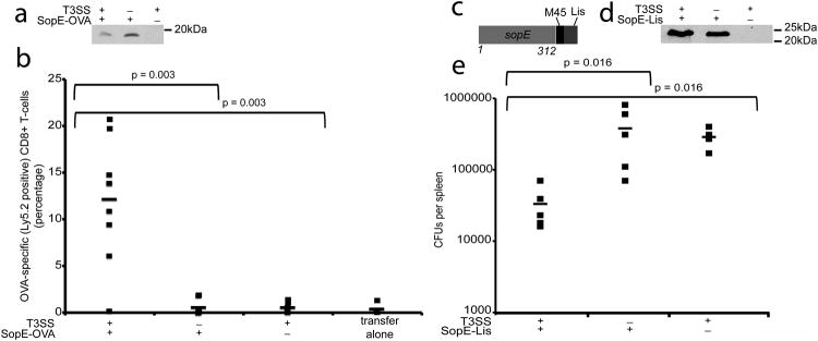
Fig. 4. T3SS-dependent priming of protective CD8+ T-cell responses by minicells.

(a) Western blot analysis of minicells obtained from wild type or T3SS-defective (ΔinvA) S. Typhimurium strains expressing the SopE-OVA construct and used in the experiment shown in (b). Equal amount of total protein was loaded in each sample. (b) Splenocytes from OT-I mice were adoptively transferred into recipient mice (C57BL/6/CD45.1), which were subsequently immunized with minicells isolated from the indicated S. Typhimurium Δasd strains expressing SopE-OVA. Three weeks after minicell immunization mice were boosted and the levels of OVA-specific CD8+ T cells were measured by flow cytometry as indicated in Materials and Methods. Values represent the percentage of OVA-specific CD8+ T-cells in each individual mouse (number of mice used in each category: T3SS+/SopE-OVA+: 9; T3SS−/SopE-OVA+: 7; T3SS+/SopE-OVA−: 7; 4 transfer alone: 4). Data were analyzed using the Student's t test. (c) Schematic of SopE-Lis construct used in the protection experiments. (d) Western blot analysis of minicells obtained from wild type or T3SS-defective (ΔinvA) S. Typhimurium strains expressing the SopE-Lis construct and used in the experiment shown in (e). (e) BMDCs prepared from Balb/c mice were incubated with minicells isolated from the indicated bacterial strains and transferred by tail vein injection into a Balb/c mouse as indicated in Materials and Methods. Six days after transfer mice were challenged with L. monocytogenes, and 3 days after challenge the c. f. u. in spleens were determined (number of mice used in each category: T3SS+/SopE-Lys+: 5; T3SS−/SopE-Lys+: 5; T3SS+/SopE-Lys−: 4). Data were analyzed using the Wicoxon rank test.
