
Figure 5.
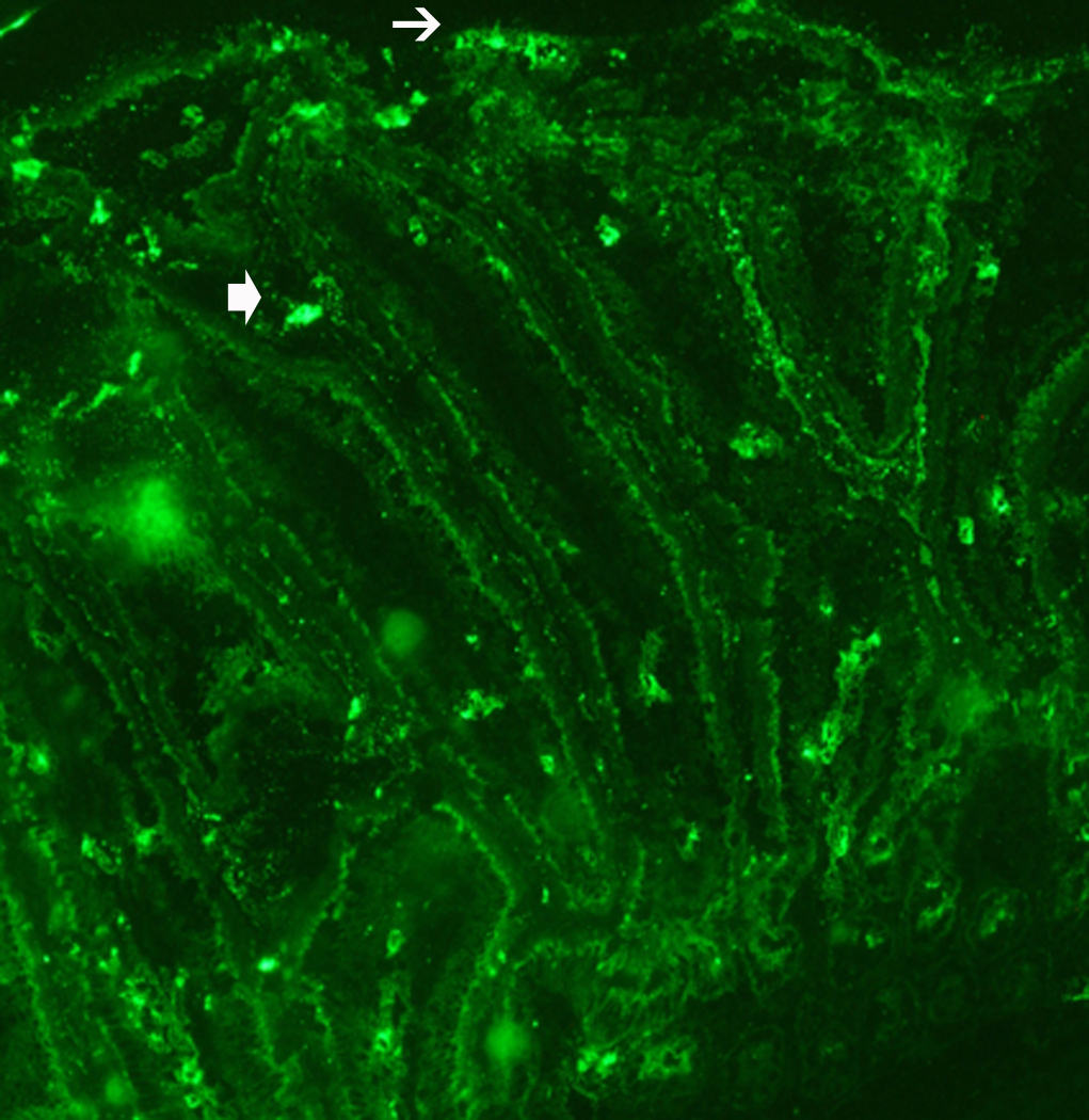
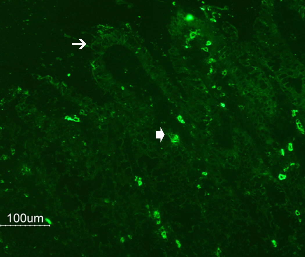


N. brasiliensis infection induced a decrease in PAR2 expression on the epithelial cell surface. (A) Frozen tissue blocks of mid-jejuna were prepared and the sections were cut for immunofluorescence staining for PAR2. The pictures are representative of each group of at least 3 mice. Original magnification, 200x. (A) Tissue from uninfected mice shows staining on epithelial cells (arrows) and on cells in the lamina propria that may be resident mast cells (arrow heads). (B) Tissue from infected animals shows diminished staining in epithelial cells (arrows) and increased staining in lamina propria cells (arrow heads). (C) A representative western blot for total PAR2 staining in scraped mucosa is shown. Total immunoreactivity was 3-fold higher (p<0.05) in infected mice, with ~80% directed to a 17 kDa form of PAR2. (D) Western blot analysis for PAR2 was performed on fractionated mucosa along with membrane or cytosolic markers (as indicated). The full size PAR2 proteins co-partitioned with plasma membrane markers (Detergent Soluble, D.S.), while the 17 kDa form was co-fractionated with cytosolic markers (Cyt), consistent with an internalization following PAR2 activation.