
Fig. 7.
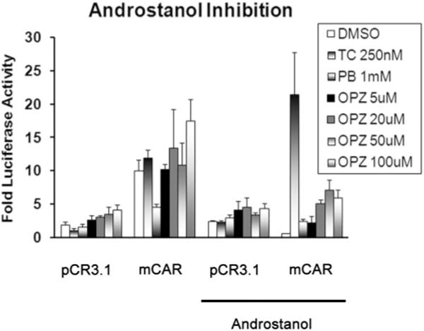
Activation of the CYP2B6-tk-luciferase reporter construct in HepG2 cells after treatment with TCPOBOP, PB, and OPZ. HepG2 cells were transiently transfected with the CYP2B6-tk-luciferase plasmid (0.1 μg) and pRL-TK (0.001 μg) into HepG2 cells, with or without a mouse CAR expression plasmid (0.25 μg). Twenty-four hours after transfection, the cells were treated with DMSO, TCPOBOP (TC, 250 nm), PB (1 mM), or OPZ (5, 20, 50, 100 μm) in the presence or absence of 10 μM 5α-androstan-3α-ol. Twenty-four hours after treatment the cell lysates were collected, and luciferase activity was determined by the Dual-Glo Luciferase Assay (Promega Corp.). The data are presented as the mean -fold activation + S.E.M. (n = 3 wells/treatment); *, significantly different from DMSO control (p ≤ 0.05).