
Fig. 1.
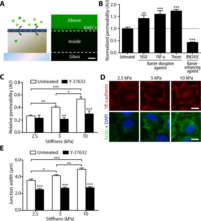
Matrix stiffness increases endothelial permeability and cell-cell junction width. (A) A cartoon shows FITC-dextran permeation through an endothelial monolayer into a polyacrylamide gel. On the right is a representative confocal cross-section of dextran capture in a 2.5-kPa polyacrylamide gel, depicting the method used to measure permeability. Dashed lines represent the top and bottom edges of the gel. Permeability values of cell (BAEC)-seeded gels were calculated as the pixel intensity within the gel divided by the pixel intensity above the gel normalized to that calculated in cell-free gels. Scale bar, 20 μm. (B) Effects of known barrier-disruptive (VEGF, TNF-α, or thrombin) and barrier-enhancing (BW245C) agonists on the permeability of BAECs cultured on 5-kPa gels. Permeability values are normalized to that of untreated cells. Data are means ± SEM. **P < 0.01, ***P < 0.001 (Dunnett’s test) compared to untreated cells. (C) Relative permeability of BAECs cultured on substrates matching (2.5 kPa) and exceeding (5 and 10 kPa) the stiffness of bovine arterial intima with (n = 8 gels, three independent experiments) or without (n = 16–20 gels, ten independent experiments) Y-27632 treatment. Data are means ± SEM. *P < 0.05, **P < 0.01, ***P < 0.001 (Tukey’s test) compared to respective untreated conditions unless otherwise indicated by brackets. (D) Fluorescent images showing VE-cadherin (red), actin (green), and nuclei (blue) of confluent BAECs on gels. (E) VE-cadherin junction-width measurements of BAECs on gels with or without 30 min Y-27632 treatment (n = 60 junction measurements, two independent experiments). Scale bar, 10 μm. Data are means ± SEM. *P < 0.05, **P < 0.01, ***P < 0.001 (Tukey’s test) compared to respective untreated conditions unless otherwise indicated by brackets.