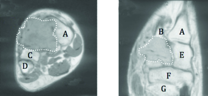
Figure 2.

Magnetic resonance imaging. a) coronal and b) axial sections through right mid foot demonstrating soft tissue mass obliterating second and third tarsometatarsal joints (delineated by dotted line). Key: A: first metatarsal, B: second metatarsal, C: third metatarsal, D: fourth metatarsal, E: medial cuneifrom, F: navicular, G: talus.
