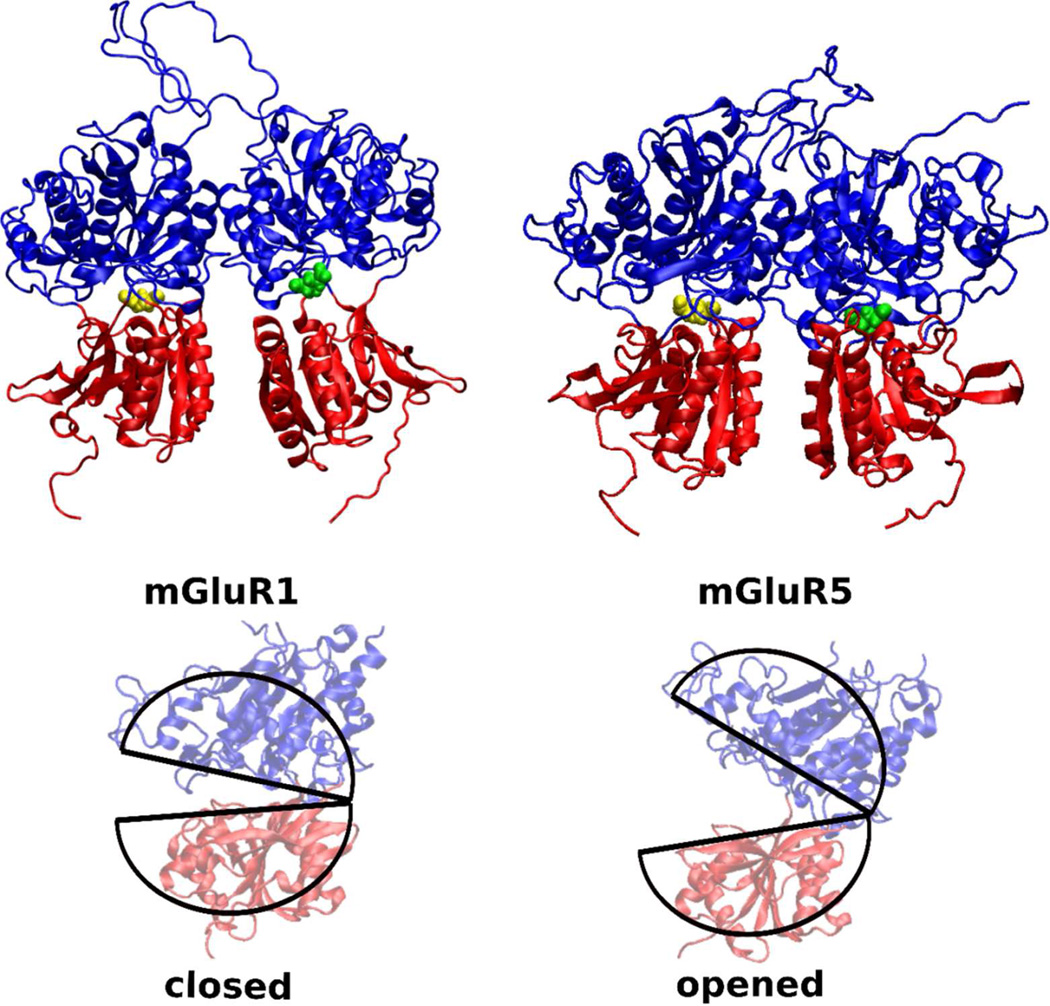
Figure 1.
The mGluR1 (PDB:1EWK) and mGluR5 (PDB:3LMK) bi-lobate structures have a strong structural similarity. The glutamate binding region is well preserved across both mGluR1 and mGluR5. Glutamate in the LBR pockets are colored in yellow and green. In mGluR1, LBR with the yellow colored glutamate represents the closed state, while the LBR with the green colored glutamate represents the open state. Both the LBRs of the mGluR5 are in the closed state. Difference in opened and closed conformatiosn of an LBR is highlighted with pie-shaped cartoon traces in the lower panel.

