Figure 1.

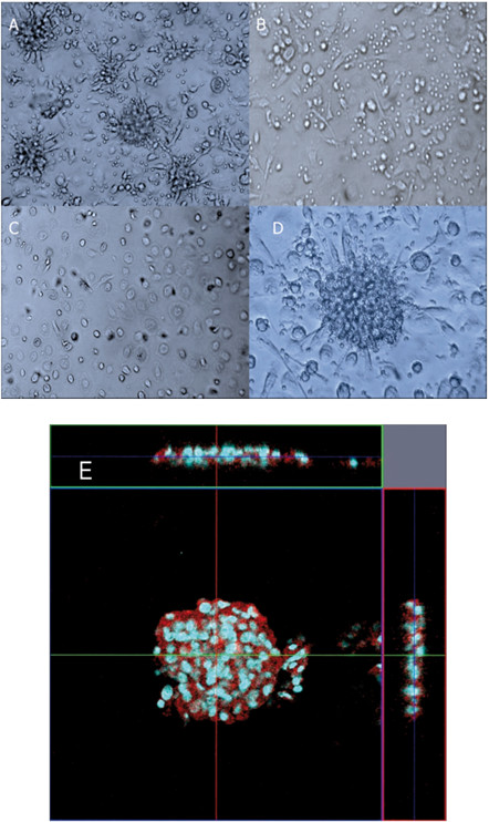
Formation of granuloma-like cellular aggregates by co-culture of PBMCs and macrophages infected with M. leprae. (A) Co-culture of macrophages (1 × 10⁵), infected with M. leprae (MOI:50) and autologous PBMCs (5 × 10⁵) in a 24 well-plate resulted in the formation of granuloma-like aggregates by day 9. (B) Culture of macrophages (1 × 10⁵) and autologous PBMCs (5 × 10⁵) for 9 days, without the bacilli. No formation of granuloma-like aggregates was observed. (C) Macrophages (1 × 10⁵) infected with M. leprae (MOI:50) after 9 days co-culture. (D) Higher magnification (2×) of the cell-aggregates in (A). (E) Confocal microscopic (LSM5 Exciter) analysis of M. leprae-induced granuloma revealed a multilayered structure (about 3–4 cell layers cells in transverse and straight sections). The cells in aggregates were positive for CD163 (red), a marker of macrophages. Nuclei were stained with Hoechst 33343 (blue). Representative data from a single donor are shown.
