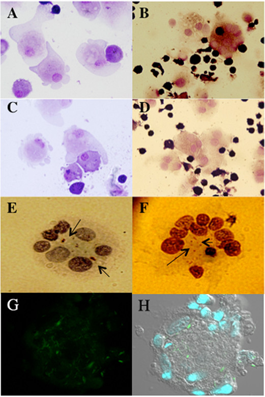
Figure 2.

Cell populations in granuloma-like aggregates. May–Grünwald–Giemsa (MGG) staining showed that there are mainly macrophages, ECs (A, D) and MGCs in the aggregates (B, D). MGCs were formed by the intercellular fusion and phagocytosis of cells (C). M. leprae were stained with Ziehl-Neelsen (shown with arrows) and the bacilli were found to be restricted to the central cytoplasmic region of the MGCs (E, F). Confocal microscopy of MGCs showed M. leprae stained with auramine O (green) and the nuclei stained with Hoescht (G, H).
