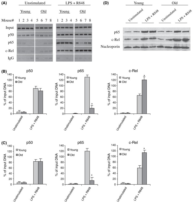
Fig. 2.
c-Rel and p65 binding to the IL-23p19 promoter in bone marrow-derived DCs from young and old mice. (A) DCs were left unstimulated or stimulated for 4 h with 100 ng mL−1 LPS plus 3 μM R848. DNA-protein complexes were cross-linked with 1% formaldehyde. Cross-linked chromatin was isolated and immunoprecipitated with c-Rel, p50, or p65 Ab. One tenth of each chromatin sample was reserved before immunoprecipitation and used as an internal control (input). The rest of the sample was used for the immunoprecipitation. DNA was recovered from the immunoprecipitation and input (10× dilution) samples and then analyzed for the presence of IL-23p19 promoter sequences by PCR. Data from four mice in each group are shown. (B) Densitometric analysis of the bands shown in (A). Band intensities were analyzed using Quantity One Imager. IgG control values were subtracted and the sample values were normalized to input DNA and are presented as percentage of input (diluted 10×). Data are the mean ± SEM and are representative of three experiments. *Statistically (P ≤ 0.05) significant (young vs. old). (C) Real-time PCR analysis of p50, p65, and c-Rel binding. Data represent the mean ± SEM of three experiments. (D) c-Rel and p65 protein expression in the nucleus of DCs. Cells were left unstimulated or stimulated for 4 h and nuclear proteins were extracted and analyzed for the expression of c-Rel and p65 by western blotting. Membranes were first probed with c-Rel Ab, stripped and re-probed with p65 and then nucleoporin Ab. Results from two mice in each group are shown.

