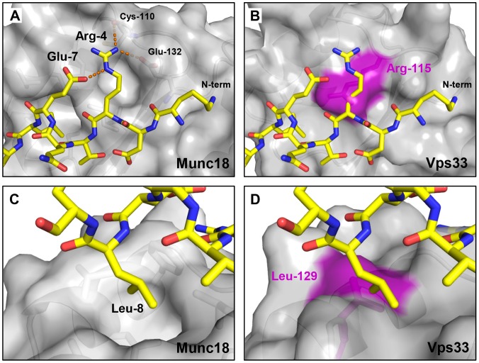
Figure 5. Alterations in Vps33 domain 1 eliminate the N-peptide binding site.
(A) Arg-4 plays a key role in the binding of the N-peptide of syntaxin 1A to domain 1 of Munc18–1 (PDB entry 3C98) [12], forming a network of hydrogen bonds and salt bridges denoted by dashed orange lines. (B) The same peptide, overlaid on the corresponding surface of Vps33, clashes with Vps33 residue Arg-115 (purple). (C) A different view of the complex shown in panel A highlights the hydrophobic pocket into which Leu-8 of syntaxin 1A packs. (D) The corresponding view of the model shown in panel B illustrates that Vps33 residue Leu-129 fills the hydrophobic residue binding pocket.

