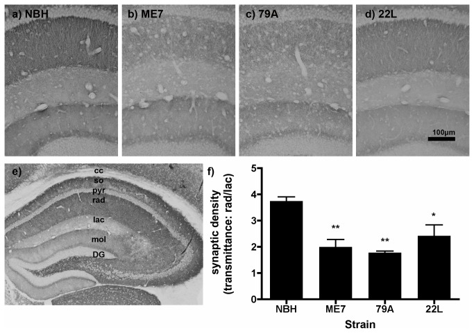
Figure 2. Hippocampal synaptophysin in prion-diseased and normal brain homogenate-inoculated animals.
Sy38 labelling of the presynaptic marker synaptophysin in a) NBH, b) ME7, c) 79A, and d) 22L at 13 weeks post-inoculation. e) The laminar structure of the hippocampus of an NBH animal: there is particularly dense synaptophysin labelling in the stratum oriens and radiatum in CA1 and CA3 and at the border between the granule cells and the molecular layer of the dentate gyrus. f) There is a significant decrease in the ratio of synaptic density in the stratum radiatum versus the synaptic density in the stratum lacunosum for all three prion strains when compared to NBH animals. ** P < 0.01, by Bonferroni post hoc test after a significant one-way ANOVA (n=3-4 sections from each of 3 animals per treatment group). Abbreviations: DG, dentate gyrus; mol, molecular layer, dentate gyrus; lac, lacunosum moleculare layer, hippocampus; so, stratum oriens; rad, stratum radiatum; pyr, pyramidal cell layer, Scale bar = 100 µm.

