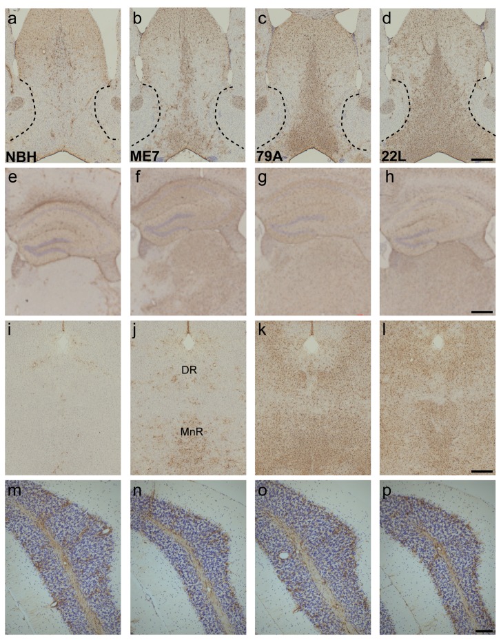
Figure 3. GFAP-positive astrocytes increase in prion-diseased and normal brain homogenate-inoculated animals.
a–d) The number of astrocytes detectable with GFAP dramatically increases in the MSDB and the dorsal part of the lateral septum of prion animals when compared to controls (3a vs. 3b-d). e–h) GFAP-positive astrocytes were present in the NBH dorsal hippocampus (3e), and were increased in all strains with robust upregulation in ME7 and 79A animals (3f & 3g) and a more variable increase in 22L animals (3h). i–l) Only blood vessel-associated astrocytes were detected in the dorsal (DR) and median raphe (MnR) in NBH animals (3i) but GFAP was readily detectable in ME7, 79A and 22L animals (3j-l). m–p) In the simple lobule of the cerebellum the scattered and variable levels of GFAP positive astrocytes were similar in all four treatment groups. Scale bars = 500 µm a-l, 100 µm m-p.

