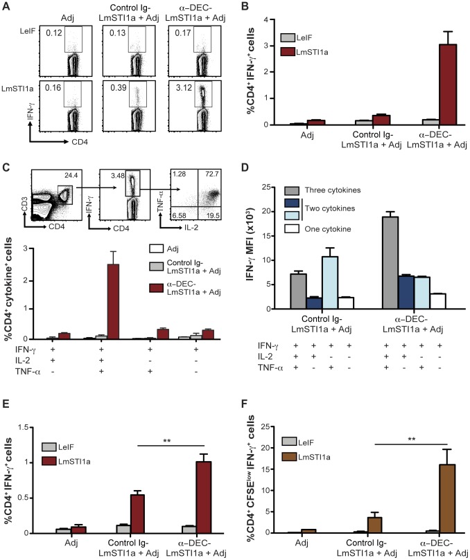
Figure 1. Single dose of anti-DEC-LmSTI1a immunizes multifunctional Th1 CD4+ T cells in vivo.
(A) C57BL/6 mice were intraperitoneally inoculated with 1 µg of anti-DEC-LmSTI1a or control Ig-LmSTI1a plus 50 µg poly ICLC and 25 µg anti-CD40 mAb (Adjuvants, Adj). Fourteen days later, splenocytes were restimulated in vitro with LmSTI1a reactive peptide mix or LeIF nonreactive peptide mix in the presence of BFA. Intracellular cytokine staining was performed to detect IFN-γ in CD3+ CD4+ T cells. (B) As in A, but % of IFN-γ+CD4+ T cells is shown as mean ± SEM (n = 9). (C) Top panel shows the gating strategy to identify multifunctional T cells using multiparameter flow cytometry. CD3+ CD4+ IFN-γ+T cells (middle FACS plot) were analyzed for production of IL-2 and TNF-α (right FACS plot). Bottom bar graph shows the frequencies of total CD4+ T cells expressing each of the four cytokine combinations. Graph is expressed as the frequency of CD4+ CD3+ T cells and the mean ± SEM (n = 9). (D) IFN-γ median fluorescence intensity (MFI) of antigen-specific CD4+ T cells producing three, two, or one cytokines (IFN-γ alone or with TNF-α and/or IL-2) following immunization of C57BL/6 mice as in A. (E) As in A, but in Balb/c mice. The % of CD4+ T cells is shown as the mean ± SEM (n = 10). (F) As in E, but bulk splenocytes were CFSE-labeled and stimulated with LmSTI1a reactive peptide mix or LeIF nonreactive peptide mix for 4 days, wherein the cells were restimulated with LmSTI1a reactive peptide mix in the presence of BFA to detect IFN-γ+ cells in proliferating CFSElow CD3+ CD4+ T cells. The frequency of CFSElow IFN-γ+ CD4+ T cells is shown as the mean ± SEM (n = 6).

