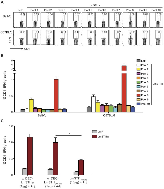
Figure 3. CD4+ T cell responses induced by anti-DEC-LmSTI1a are directed to several epitopes.
(A) Balb/c or C57BL/6 mice were intraperitoneally immunized with 1 µg of anti-DEC-LmSTI1a plus poly ICLC (50 µg) and anti-CD40 mAb (25 µg) (Adj). Fourteen days later, splenocytes were restimulated with 10 different LmSTI1a peptide pools (2 µg/ml), each containing 12 individual peptides. IFN-γ-producing CD4+ T cells were assessed by intracellular cytokine staining. (B) As in A, but the percentage of IFN-γ+ CD4+ T cells is shown as mean ± SEM (n = 6). (C) Balb/c mice were immunized with 1 µg of anti-DEC-LmSTI1a or anti-DEC-LmSTI1344–366 mAbs or 10 µg of non-targeted LmSTI1348–362 immunodominant peptide in the presence of 50 µg poly ICLC and 25 µg anti-CD40 mAb (Adj). Production of IFN-γ was evaluated by FACS after restimulation with LmSTI1a reactive peptide mix or LeIF nonreactive peptide mix. The % of IFN-γ+ CD4+ T cells is shown as mean ± SEM (n = 3).

