
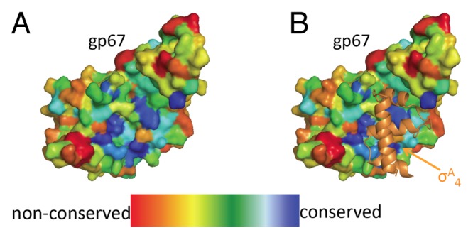
Figure 2. Structural conservation of gp67. (A) gp67 from the 2.0 Å co-crystal structure with σA4 colored by conservation. The structural conservation map was made using ConSurf using five available gp67 sequences. Highly conserved residues are shown in blue and poorly conserved residues in red. (B) Highly conserved gp67 residues map to the σA4 binding site. gp67 from the 2.0 Å co-crystal structure is shown as in (A) and σA4 from the co-crystal structure is shown in orange as a cartoon representation. The most highly conserved surface residues in gp67 map to the interaction between gp67 and σA4.