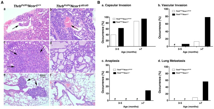
Figure 2. Expression of NCOR1ΔID delays thyroid cancer progression in ThrbPV/PVNcor1ΔID/ΔID mice.
(A) Hematoxylin and eosin (H&E) staining of thyroid sections (top and middle rows) and lung sections (bottom row) of ThrbPV/PVNcor1+/+ (a, c, and e) and ThrbPV/PVNcor1ΔID/ΔID (b, d, and f) mice. Histological sections from tissues of ThrbPV/PVNcor1+/+ mice showed evidence of (a) vascular invasion in thyroid (arrow), (c) anaplasia in thyroid (arrows), and (e) metastatic lesions in lung (arrows). Sections of thyroids and lungs from ThrbPV/PVNcor1ΔID/ΔID showed blood vessels (b) without vascular invasion (arrow), (d) without anaplasia, and (f) lung without metastatic lesions. (B) Comparison of age-dependent percentage occurrence of capsular invasion (a), vascular invasion (b), anaplasia (c), and lung metastasis (d). The data are expressed as the percentage of occurrence of total mutant mice examined. The symbol “#” indicates 0% occurrence.

