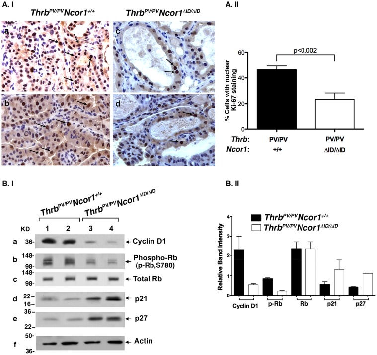
Figure 3. Expression of NCOR1ΔID inhibits proliferation of thyroid tumor cells of ThrbPV/PVNcor1ΔID/ΔID mice.
(A.I) Two representative microphotographs of stained Ki-67 on thyroid sections of ThrbPV/PVNcor1+/+ (a and b represent two different mice) and ThrbPV/PVNcor1ΔID/ΔID mice (c and d represent two different mice). In the thyroids of ThrbPV/PVNcor1ΔID/ΔID mice, fewer thyroid cells were stained with Ki-67 than in thyroids of ThrbPV/PVNcor1+/+ mice (arrows). (A.II) Positively nuclear Ki-67-stained thyroid cells from ThrbPV/PVNcor1ΔID/ΔID and ThrbPV/PVNcor1+/+ mice were counted and expressed as percentage of total cells. The percentage of proliferating cells was significantly lower in the thyroids of ThrbPV/PVNcor1ΔID/ΔID than ThrbPV/PVNcor1+/+ mice. (B.I) Nuclear protein extracts were prepared from thyroid tumors of ThrbPV/PVNcor1+/+ (lanes 1 and 2) or ThrbPV/PVNcor1ΔID/ΔID (lanes 3 and 4) mice. Western blot analysis of cyclin D1, phosphorylated Rb, total Rb, p21, and p27 are as marked, and actin (panel f) was used as loading control. (B.II) Quantitation of band intensities shown in (B.I). The protein abundances of cyclin D1 and phosphorylated Rb were lower in the thyroids of ThrbPV/PVNcor1ΔID/ΔID mice, whereas the protein abundance of p21 and p27 were higher in the thyroids of ThrbPV/PVNcor1ΔID/ΔID mice.

