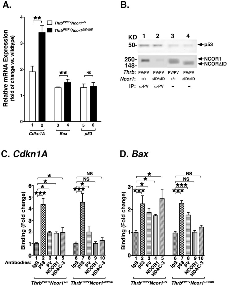
Figure 5. Expression of NCOR1ΔID alters expression of key regulators in cell proliferation and apoptosis in the thyroid of ThrbPV/PVNcor1ΔID/ΔID mice.
(A) Activated expression of the Cdkn1A and the Bax genes in the thyroid of ThrbPV/PVNcor1ΔID/ΔID mice. Quantitative real-time RT-PCR was carried out as described in Methods and Materials. Each thyroid sample was run in triplicates with total mouse numbers of 3–5. The differences in the expression are significant in the expression of the Cdkn1A (lanes 1 and 2) and the Bax genes (lanes 3 and 4) between ThrbPV/PVNcor1+/+ and ThrbPV/PVNcor1ΔID/ΔID mice (lanes 5 and 6). (B) Co-immunoprecipitation of PV with p53 and NCOR1 in the thyroid of ThrbPV/PVNcor1+/+ mice (lane 1), but not with NCOR1ΔID in thyroid extracts of ThrbPV/PVNcor1ΔID/ΔID mice (lane 2). Lanes 3 and 4 show the p53 band (upper panel) and NCOR1 and NCOR1ΔID from direct Western blot analysis from the thyroid nuclear extracts of ThrbPV/PVNcor1+/+ and ThrbPV/PVNcor1ΔID/ΔID mice, respectively. (C and D) Recruitment of PV and NCOR1 to the p53/DNA binding sites on the promoter of the Cdkn1A gene (C) and the Bax gene (D). ChIP assay was carried out using IgG (bars 1 and 6) or anti-p53 (bars 2 and 7) or anti-PV (bars 3 and 8) or anti-NCOR1 (bars 4 and 9) or anti-HDAC-3 (bars 5 and 10) antibodies as described in Methods and Materials. Binding was expressed as fold of changes in reference to the negative control in which mouse IgG was used in the immunoprecipitation.*p<0.05, and ***p<0.001. NS, not significant.

