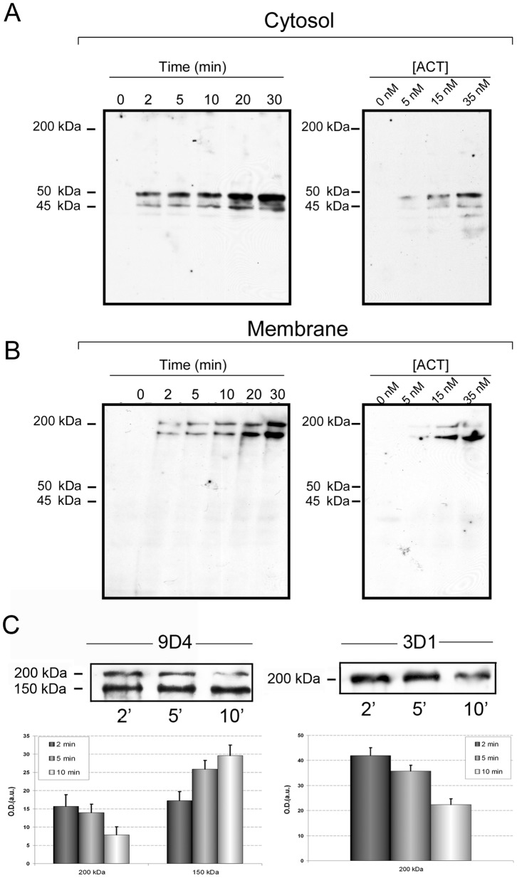
Figure 1. ACT is proteolytically cleaved in J774A.1 cells.
Western blot analysis of cytosolic extracts (A) and membrane fractions (B) purified, as described in Materials and Methods, from J774A.1 cells treated with ACT (35 nM) at different incubation times (0–30 min) or different toxin concentrations (0–35 nM), and stained with anti-AC monoclonal antibody MAb 3D1 for the cytosolic fraction, and with MAb 9D4 for the membrane fraction. (C) Time course of the 200 and 150 kDa bands in membrane fractions of cells treated with 5 nM ACT at different incubation times. J774A.1 cells were incubated with ACT at 4°C and non-bound ACT was washed in ice-cold PBS as described in Materials and Methods. Then, cells were challenged at 37°C and the full length toxin and the 150 kDa fragment were determined in the membrane fractions of the cells with MAb 9D4 (left-hand panel) and the full length toxin was also detected by MAb 3D1 (right-hand panel). Densitometric quantification of the bands detected in the immunoblots is shown below the Western blots. A representative experiment from three independently performed assays is shown in (A) and (B), and a representative experiment from two independently performed assays is shown in (C).

