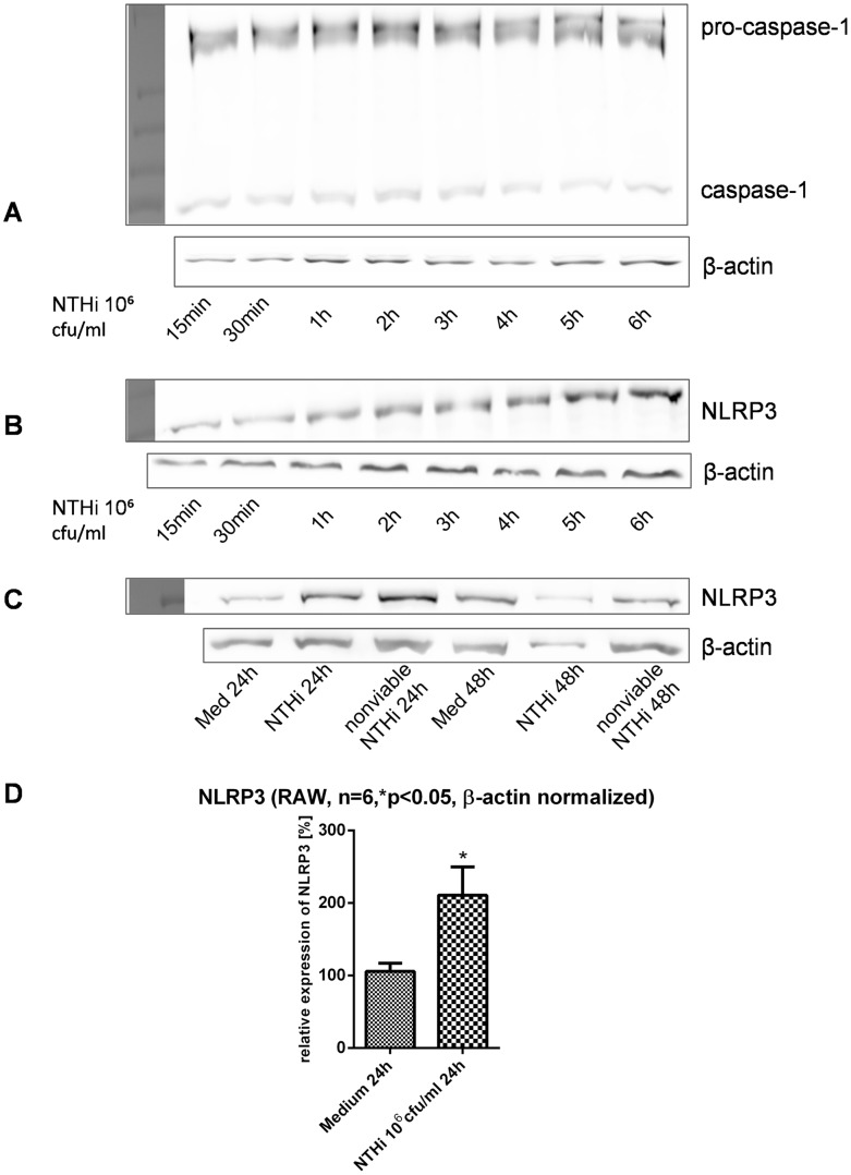
Figure 3. Western Blot analysis of inflammasome components after stimulation with NTHi.
A+B show caspase-1 (A) and NLRP3 (B) expression over a period of 6 h after stimulation with NTHi 106 cfu/ml in murine macrophages. NLRP3 was detected in murine macrophages after stimulation with inactivated NTHi as well (C). Densitometric analysis of NLRP3 in murine macrophages after stimulation with NTHi 106 cfu/ml for 24 h is shown in (D).

