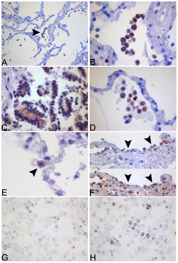
Figure 4. Immunohistochemical and immunocytochemical staining for NLRP3 in human lung tissue and human alveolar macrophages.
Human lung tissue was stimulated with NTHi 106 cfu/ml for 24 h. Tissue was fixated with HOPE-solution and NLRP3 detected via IHC. The images show the expression of NLRP3 in alveolar macrophages (A+B), bronchial epithelial cells (C), in the medium control (D) and in alveolar type II cells (ATII) (E). A specific ATII staining (human surfactant protein B) was performed in HLT (F, upper image). NLRP3-positive cells (F, lower image) were identified in consecutive slides from the same tissue section (arrows). Human alveolar macrophages from BAL were stained for NLRP3. Increased expression can be observed after stimulation with NTHi (H) in contrast to the medium control (G).

