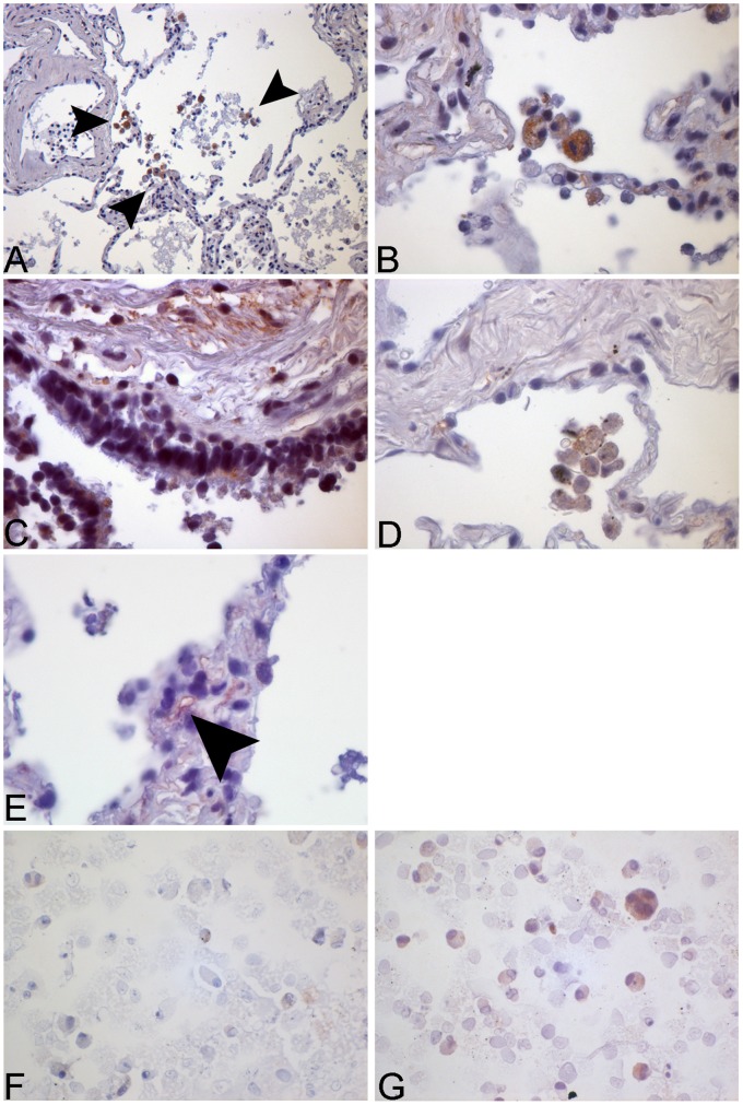
Figure 5. Immunohistochemical and immunocytochemical staining for caspase-1 in human lung tissue and human alveolar macrophages.
Human lung tissue was stimulated with NTHi 106 cfu/ml for 24 h. Tissue was fixated with HOPE-solution and caspase-1 detected via IHC. The images show the expression of caspase-1 in alveolar macrophages (A+B), bronchial epithelial cells (C), in the medium control (D) and in the capillary endothelium (arrow) (E). Human alveolar macrophages from BAL were stained for caspase-1. Increased expression can be observed after stimulation with NTHi (G) in contrast to the medium control (F).

