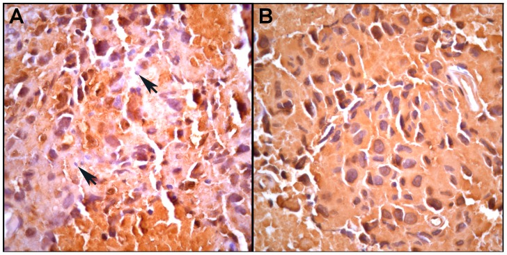
Figure 2. An adjacent section to that in Fig. 1 stained for the melanoma-specific antigen S100.
A. An area of S100-positive tumor cells admixed with infiltrating S100-negative leucocytes (arrows). B. An area containing only S100-positive tumor cells. More detailed pathology analyses are presented in Figs S3 and S4 in File S1.

