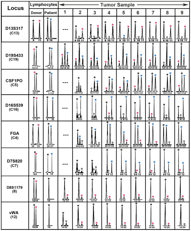
Figure 3. Forensic STR analyses of the MH3 melanoma along with donor and patient pre-BMT lymphocytes.
Shown are “informative” loci exhibiting donor and patient specific alleles in pre-BMT lymphocytes. Tumor loci are listed in order of relative abundance of the donor-specific alleles (red asterisk) compared to patient-specific (blue asterisk) and shared alleles (black asterisk). Allele peaks <50 relative fluorescence units were censored as “no call” (open circles). Loci with no detectable alleles after PCR amplification (—).

