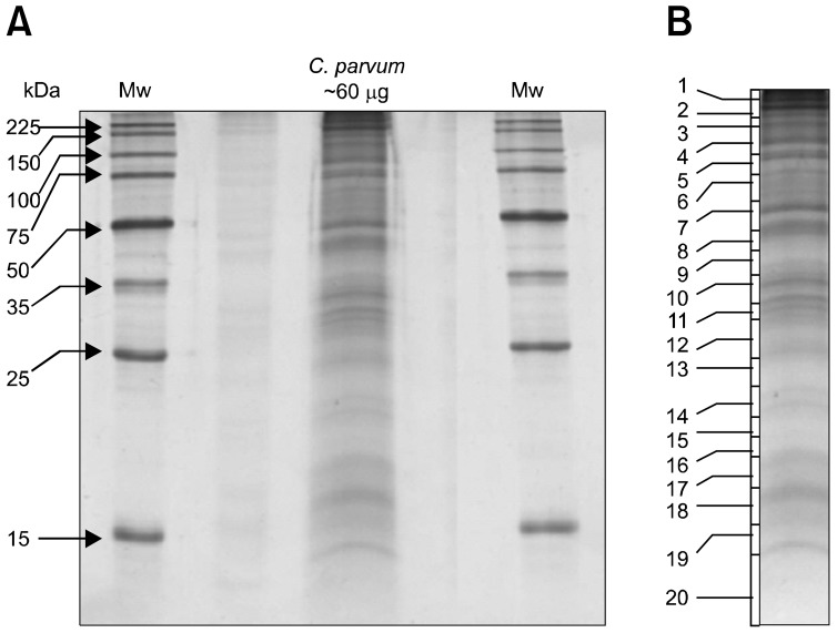
Fig. 1.
(A) 1D-SDS-PAGE analysis of sporozoite proteins of Cryptosporidium (C.) parvum. Total no. of oocysts ~ 107. Total amount of protein~60 µg. Electrophoresed proteins were visualized with colloidal coomassie stain. Molecular weight markers (in kilodaltons) are shown on the left. (B) The lane containing C. parvum proteins was excised into 20 slices. Each gel slice was then digested by trypsin and analyzed by LC-MS/MS.

