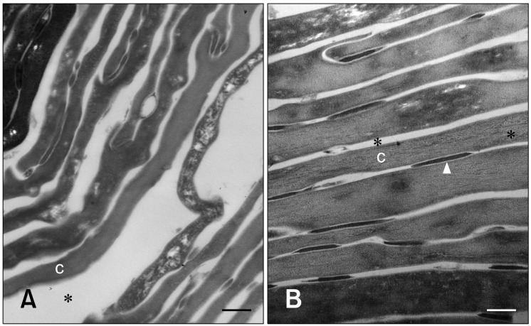
Fig. 2.
Transmission electron micrographs of the upper epidermis before and after moisturizer treatment. (A) Before treatment, corneocytes were arranged in a disorganized manner and were less compact with wide intercellular spaces. (B) After moisturizer treatment, corneocyte organization was more regular and compact in the stratum corneum. Ruthenium tetroxide post-fixation. Asterisks (*) indicate intercellular space. Arrowhead: desmosome, C: corneocyte. Scale bars = 200 nm.

