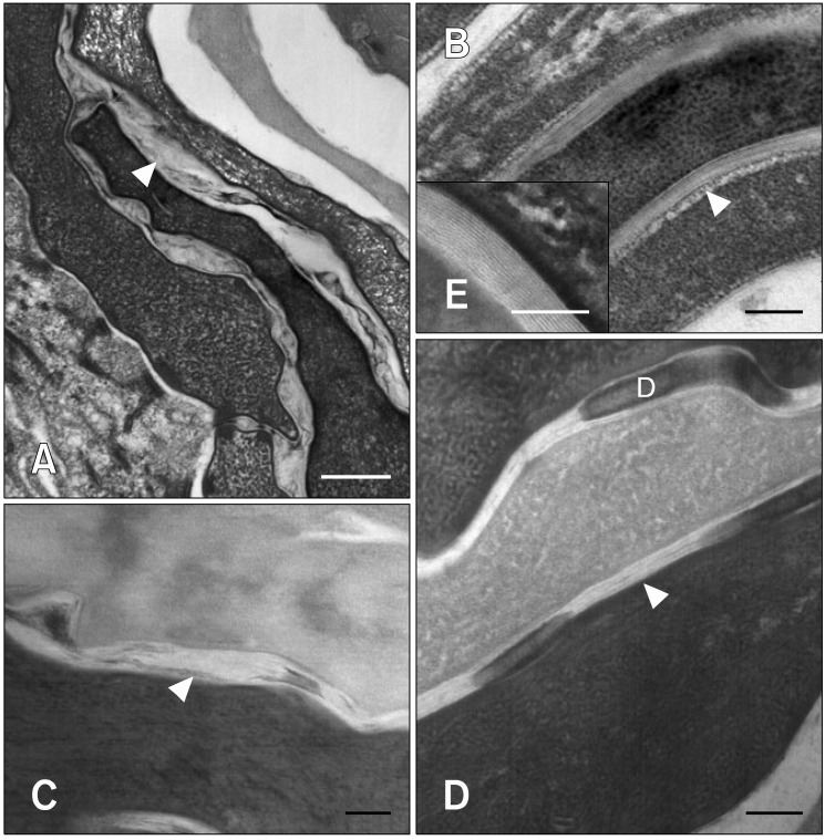
Fig. 3.
Transmission electron micrographs of the stratum corneum before and after moisturizer treatment. (A, C) In atopic dogs, the lipid lamellae were greatly disorganized and the reduced intercorneocyte space was occupied by lipid lamellae. (B, D) After moisturizer treatment, the lipid lamellae were more organized and the increased intercorneocyte space was occupied by a nearly normal lipid bilayer. (E) Magnified field from Fig. 3B. The lipid bilayer was composed of alternating layers of electron-dense lamellae and electron-lucent lamellae. Ruthenium tetroxide post-fixation. Arrowheads: lipid lamellae, D: desmosome. Scale bars = 100 nm.

