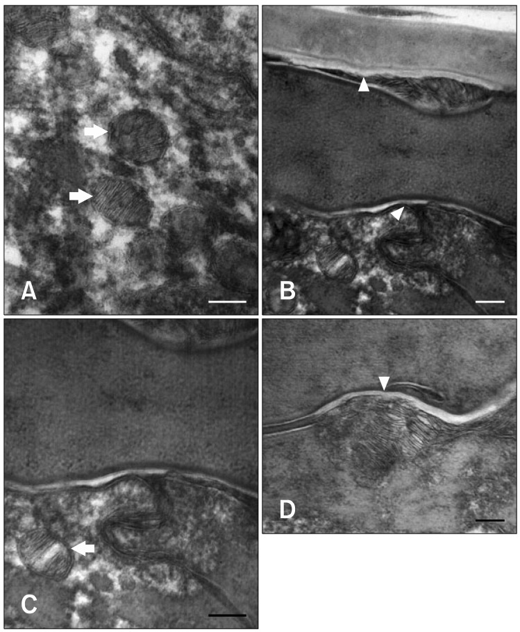
Fig. 4.
Transmission electron micrographs of the interface between the stratum corneum and stratum granulosum after moisturizer treatment. Following moisturizer treatment, more active lamellar body extrusion was observed. (A) Lamellar body structures in the stratum granulosum. (B, C and D) Secretion of lamellar body lipids and their transformation into the lipid bilayer. After extrusion of the lamellar body disks into the intercellular space, the brims of the adjacent disks fused and formed continuous lipid bilayers. Ruthenium tetroxide post-fixation. Arrows: lamellar body, Arrowheads: lipid bilayer. Scale bars = 100 nm.

