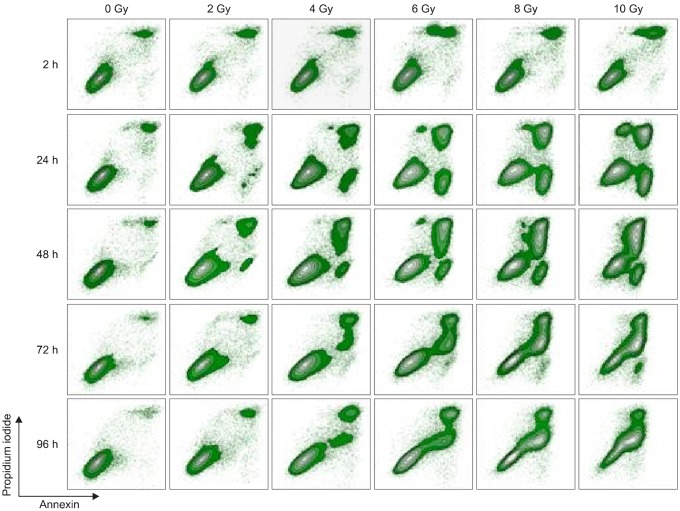
Fig. 5.
Radiation induced apoptosis of TLM 1 cells analyzed by FCM (annexin/PI iodide staining). Cells were gated according to FSC/SSC light scattering properties (not depicted). Results are displayed in contour plots. One representative sample of three is shown. FCM: flow cytometry, PI: propidium iodide, FSC/SSC: forward scatter/side scatter.

