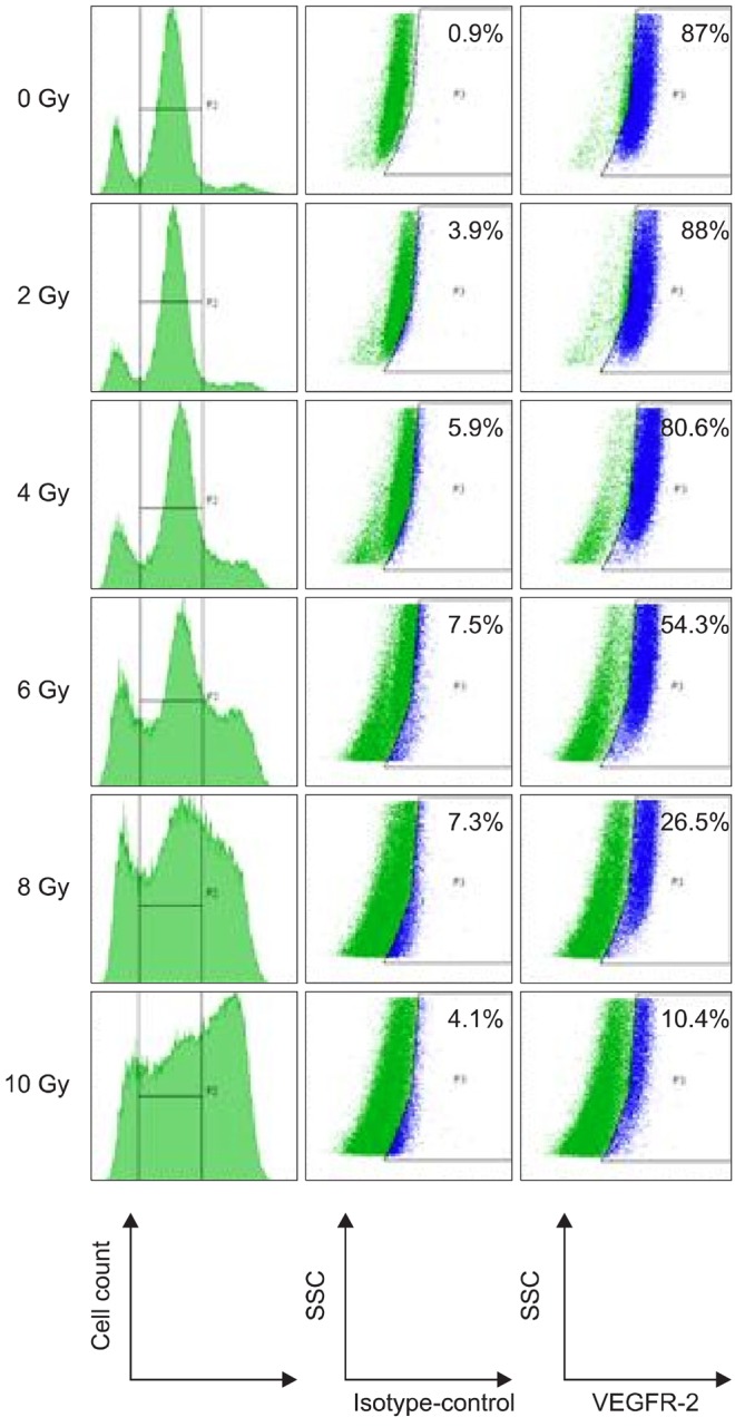
Fig. 6.

FCM analysis of VEGFR-2 in cytoplasm of TLM 1 cells four days post radiation (0, 2, 4, 6, 8 and 10 Gy). Cells were stained with live/dead stain, after which they were fixed, permeabilzed and stained with antibodies against VEGFR-2 or irrelevant isotype control antibodies. Only live cells were gated and analyzed for VEGFR-2 expression. VEGFR-2 positive cells are highlighted in blue. The percentage of VEGFR-2 positive cells is shown in the upper right corner. VEGFR: vascular endothelial growth factor receptor.
