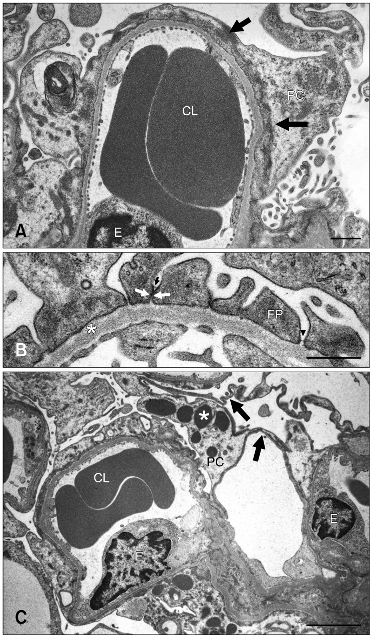
Fig. 2.
(A) Severe podocyte foot process (FP) effacement with rearrangement of the actin cytoskeleton (arrows) over broad areas of the glomerular basement membrane (GBM) in a 9-week old male OM rat. (B) FPs anchoring the GBM (*) in close contact with one another or adhered (arrows) with dislocation of the slit membrane in a 6-week old OM rat. Arrowhead indicates the slit membrane. (C) Osmiophilic lysosomes (*) and flattened podocytes (arrows) in a 24-week old male OM rat. CL: capillary lumen, E: endothelial cell, FP: foot process, PC: podocyte. Scale bars = 1 µm (A and B) and 3 µm (C).

