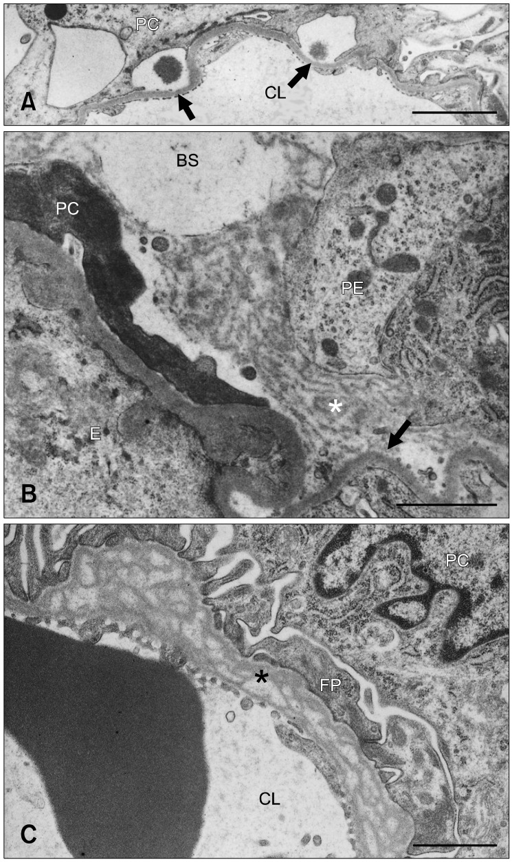
Fig. 3.
(A) Denuded GBM (arrows) resulting from FP detachment in a 24-week old male OM rat. (B) An electron-dense matrix (*) formed a bridge between the denuded GBM (arrow) and parietal epithelial cells rich in polysomes and rough endoplasmic reticula in a 24-week old male OM rat. A degenerated podocyte is shown on the left side. (C) Prominent splitting or lamination of the GBM (*) in a 6-week old male OM rat. BS: Bowman's space, CL: capillary lumen, E: endothelial cell, FP: foot process, PC: podocyte, PE: parietal epithelial cell. Scale bars = 3 µm (A) and 1 µm (B and C).

