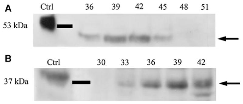
Fig. 2.
The proteolytically processed fragments of DMP1 in mouse SMG. Ten 12-week-old mice were used to extract proteins and then perform western immunoblotting. a Western immunoblotting for the C-terminal fragment of DMP1 in mouse SMG using the polyclonal antibody anti-DMP1-C-857. Positive control (Ctrl) was 1 μg of C-terminal fragment of DMP1 isolated from the rat incisors, which migrated at 57 kDa. Approximately 46 kDa protein band representing the C-terminal fragment of DMP1 (arrow) was observed in fractions 36–45 of the Q-Sepharose chromatography. b Western immunoblotting for the N-terminal fragment in mouse SMG using the monoclonal antibody anti-DMP1-N-9B6.3. Positive control (Ctrl) was 1 μg of DMP1 isolated from rat incisors. The 37 kDa N-terminal fragment of DMP1 (arrow) was detected in fractions 33–42

