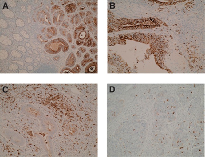
Figure 1.
Expression of NGAL in colorectal neoplastic progression. (A) NGAL epithelial and stromal cell staining in the adenomatous compartment of a CaP lesion, with no positive staining seen within the normal mucosa ( × 20 magnification). There is a clear demarcation in staining pattern between these compartments highlighting that NGAL expression occurs early in the adenoma–carcinoma sequence with a sharp transition between normal and dysplastic epithelium. (B) Epithelial and stromal cell NGAL positivity within colonic adenoma exhibiting low-grade dysplasia ( × 40 magnification). (C, D) Epithelial and stromal cell NGAL positivity within carcinomatous epithelial compartment of a CaP lesion ( × 40 magnification).

