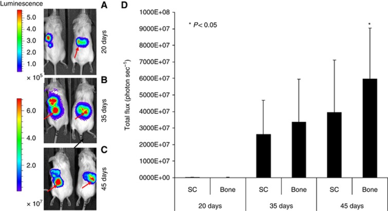
Figure 1.
Bioluminescence imaging (BLI). (A) Tumour growth was examined by BLI. Luc-CSCs-like were injected SC into flank of the SCID mice. Representative images of two animals after different time points (20, 35, and 45 days after SC injection) are shown. (B) At day 20, the SC tumour mass is clearly evident, whereas the bone localisation is detectable only in one mice (arrow). (C) At day 35, an increased tumour volume and invasion to human bone becomes particularly evident (arrow). (D) At 45 days, the signal derived from bone localisation of the CSCs-like is stronger in bone than that in SC tumour mass.

