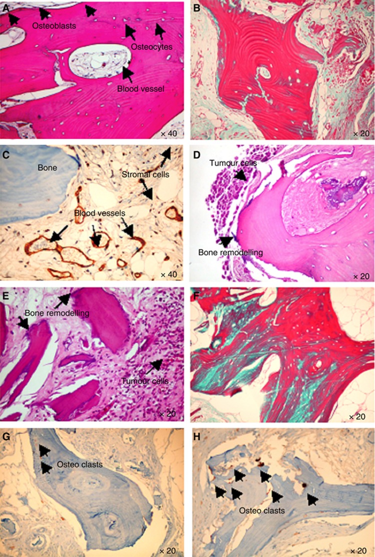
Figure 2.
Histological analysis of the implanted bone. (A) H&E-stained section in control mice shows the presence of human live bone, blood vessels, newly synthesised bone with osteoblasts lining cells, as indicated by the arrows (magnification × 40). (B) Trichrome staining shows in blue the new collagen fibres in the bone: osteoid originated after the bone implanted in SC of the control mice. (C) Human endothelial cells in the blood vessels are stained for anti-human CD34, as indicated by the arrows. (D and E, respectively) Breast Luc-CSCs-like metastasise the human-implanted bone after IC and SC injection, in both cases area of pathological bone resorption are evident, as indicated by the arrows. (F) A marked neo-bone apposition is evident (osteoid is indicated by the blue staining) in bone invaded by breast CSCs-like. Osteoclasts stained for TRAP in bone of control mice (G) and mice injected with breast CSCs-like (H).

