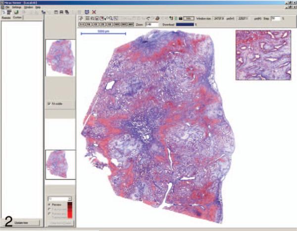
Figure 2.
The image shown in the Mirax viewer screen is a trichrome stain of one of the discrepant cases interpreted as biliary adenofibroma by the whole-slide reviewer and mesenchymal hamartoma by the glass-slide and reference reviewers. The main image can be magnified and moved about the screen by the use of a mouse and/or function buttons above the image.

