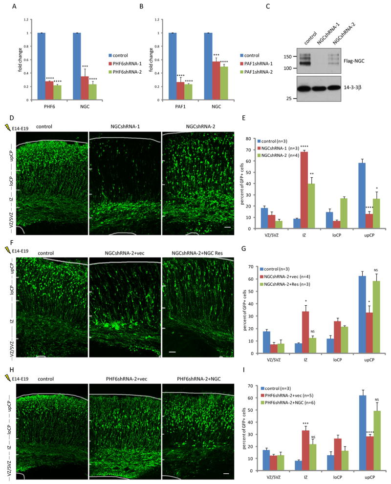
Fig. 4. The Neuroglycan C/Chondroitin sulfate proteoglycan 5 (NGC/CSPG5) gene is a key downstream target of PHF6 and PAF1 in the control of neuronal migration in vivo.
(A) Quantitative RT-PCR analyses of primary rat cortical neurons infected with lentivirus expressing PHF6 shRNAs or control shRNAs. Levels of PHF6 and NGC/CSPG5 were normalized to GAPDH levels (n=3). (B) Quantitative RT-PCR analyses of primary rat cortical neurons infected with lentivirus expressing PAF1 shRNAs or control shRNAs. Levels of PAF1 and NGC/CSPG5 were normalized to GAPDH levels (n=3). (C) Lysates of 293T cells transfected with an expression vector encoding FLAG-NGC/CSPG5 together with an RNAi plasmid encoding NGC/CSPG5 shRNAs or control shRNAs were immunoblotted with the FLAG and 14-3-3β antibodies. (D) E14 mouse embryos were electroporated with an RNAi plasmid encoding NGC/CSPG5 shRNAs or control shRNAs and analyzed as in Figure 1D. (E) Quantification of the percentage of GFP positive neurons in distinct regions of the cerebral cortex in E19 mouse embryos treated as in (D) is presented as mean + SEM. Knockdown of NGC/CSPG5 also inhibited proliferation of cortical precursor cells (see Figure S2F). (F) E14 mouse embryos were electroporated with the NGC/CSPG5 RNAi or control RNAi plasmid together with an expression plasmid encoding NGC-Res or its corresponding control vector, and analyzed as in (D). (G) Quantification of the percentage of GFP positive neurons in distinct regions of the cerebral cortex in E19 mouse embryos treated as in (F) is presented as mean + SEM. Expression of NGC-Res significantly increased the percentage of neurons reaching the upper cortical plate in the background of NGC/CSPG5 RNAi in the cerebral cortex in vivo (ANOVA, p<0.05). (H) E14 mouse embryos were electroporated with the PHF6 RNAi or control RNAi plasmid together with an expression plasmid encoding NGC/CSPG5 or its corresponding control vector, and analyzed as in (D). (G) Quantification of the percentage of GFP positive neurons in distinct regions of the cerebral cortex in E19 mouse embryos treated as in (H) is presented as mean + SEM. Expression of NGC/CSPG5 significantly increased the percentage of neurons reaching the upper cortical plate in the background of PHF6 RNAi in the cerebral cortex in vivo (ANOVA, p<0.01). See also Figure S2 and Table S1. Scale bars: 50μm in (D), (F) and (G).

