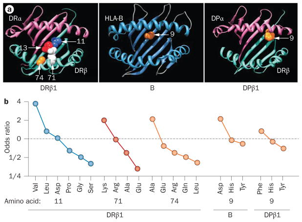
Figure 1.
Antigen-binding groove HLA amino acid substitutions and influence on susceptibility to RA. a | Three-dimensional ribbon models for the MHC class I molecule HLA-B and for the MHC class II molecules HLA-DRβ1 and HLA-DPβ1. Direct views of the peptide-binding groove are presented, showing key amino acid positions identified in an association analysis by Raychaudhuri, S. et al. Nat. Genet. 44, 291–296 (2012).38 © NPG b | The odds ratio for association with RA depends on which amino acid is substituted at positions 11, 71 and 74 of HLA-DRβ1, at position 9 of HLA-B or at position 9 of HLA-DPβ1. Abbreviation: RA, rheumatoid arthritis.

