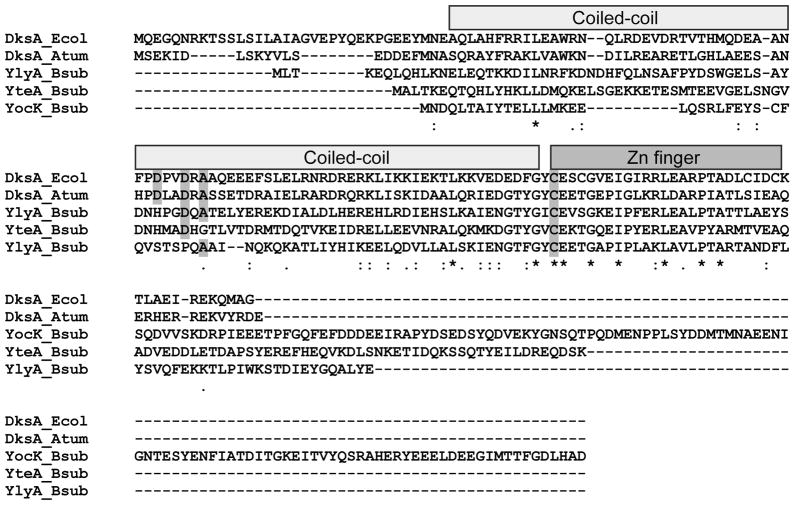
Fig. 1. Alignment of the amino acid sequences of DksA-like proteins.
ClustalW was used to align the amino acid sequence of E. coli and A. tumefaciens DksA, and B. subtilis YlyA, YocK and YteA. Identical residues (*), conserved substitutions of residues with similar properties (:), and semi-conserved substitutions of residues with similar steric confirmations (.) are indicated below the alignment. The N-terminal α-helix coiled-coil (light grey) and the C-terminal Cys-4 zinc finger motif (dark grey) are indicated above the alignment. The conserved residues of the DxxDxA motif, and the conserved first cysteine residue of the Cys-4 zinc finger motif are shaded in grey.

