
Fig. 3. Protein Level of 70S subunit from the ΔL21 mutant ribosomes.
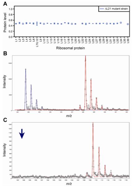
(a) Protein level for a single fraction from the 70S subunit peak. Error bars represent the standard deviation from multiple measurements from different peptides/ions in the same protein. In the case were a single measurement was obtained, an open circle is used. (b) A peptide from L5 (MWEFFER) is identified as being present in both the L21 mutant strain (blue) and the 15N ribosomal standard (red). (c) A peptide from L21 (MYAVFQSGGK) is identified as being present in the 15N ribosomal standard (red) but absent in the L21 mutant strain. The position of where the 14N peptide would appear is shown with a blue arrow.