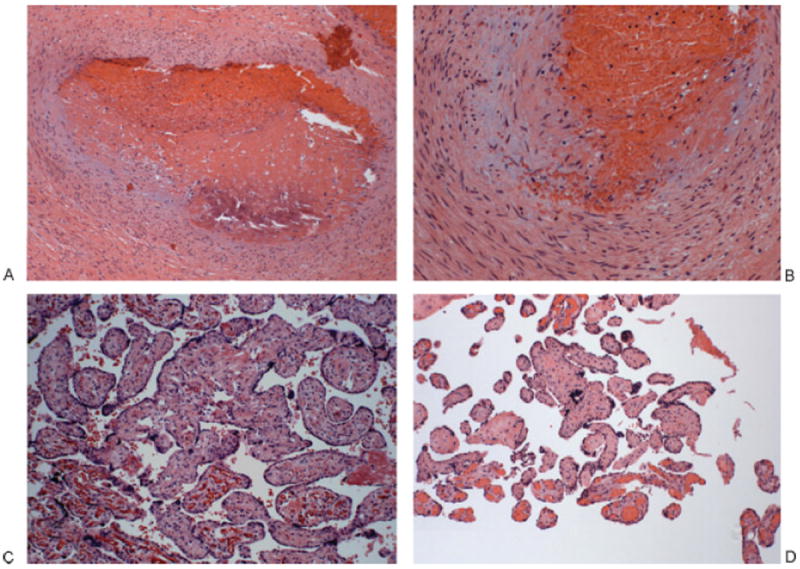
Figure 1.

Placenta of patient 1. (A, B) Thrombosed chorionic plate vessel: The vessel lumen is obliterated, filled with an organized, partially calcified thrombus (left); higher power shows a damaged vessel wall with fibrin and extravasated red blood cells; the endothelium is unrecognizable. (A) 100×. (B) 200×. (C) Terminal chorionic villi with villous stromal karyorrhexis. The villi on lower left panel are normal with intact fetal vessels. The remainder shows partially obliterated fetal vessels and a fibrotic stroma with extravasated red cells and karyorrhectic debris. 200×. (D) Terminal chorionic villi, completely avascular. The villi at the bottom are normal with intact fetal vessels; the remainders are completely avascular, with fibrosis. 100×.
